Patello-Femoral Instability
Patient information — Dr Christopher Spelman, Orthopaedic Surgeon
Overview
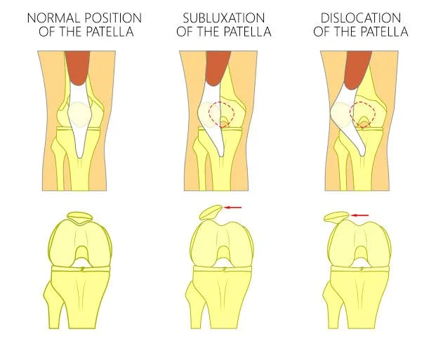
Patellofemoral instability describes a kneecap (patella) that slips partially (subluxation) or completely (dislocation) out of the groove (trochlea) at the front of the thigh bone, almost always towards the outer side. It may occur as a one-off injury from a direct blow or awkward twist, or it may become recurrent, where the kneecap dislocates repeatedly with progressively less force. Recurrent instability can damage the cartilage on the back of the kneecap and lead to long-term pain and arthritis, so it is important to understand and address the underlying causes.
Why patellofemoral instability is multifactorial
The kneecap is kept in its groove by a balance of bony shape, ligaments, muscle control and the overall alignment and rotation of the leg. Instability is rarely due to a single cause – in most patients several factors combine, and stability fails when enough of them are present. Successful treatment depends on identifying which factors are contributing in each individual. The main factors are:
Trochlear dysplasia – a shallow, flat or dome-shaped groove that does not hold the kneecap securely. This is one of the most important bony risk factors.
Patella alta – a kneecap that sits too high, so it does not engage the groove until the knee is well bent, leaving it unstable in early bending.
Increased tibial tubercle–trochlear groove (TT–TG) distance – the point where the kneecap tendon attaches to the shin is positioned too far to the outer side, pulling the kneecap laterally.
Increased femoral anteversion (torsion) – excessive inward twist of the thigh bone, which rotates the groove and directs the pull of the muscles unfavourably.
External tibial torsion and knock-kneed (valgus) alignment – rotational and coronal alignment of the leg that increases the outward pull on the kneecap.
Medial patellofemoral ligament (MPFL) insufficiency – the main soft-tissue checkrein on the inner side is torn or stretched during a dislocation, removing the primary restraint.
Generalised ligamentous laxity – naturally loose tissues, and weakness or poor timing of the quadriceps (particularly the inner portion) and hip muscles.
Assessment
Because the causes are multiple, assessment is detailed. Dr Spelman will examine the knee and the alignment and rotation of the whole limb, and will usually obtain imaging. X-rays and, importantly, cross-sectional imaging (CT or MRI) are used to measure the depth of the groove, the height of the kneecap, the TT–TG distance and the rotation of the thigh bone, and to check for cartilage or loose-fragment damage. This “map” of the contributing factors guides treatment.
Non-operative treatment
After a first-time dislocation, most patients are managed without surgery. Treatment focuses on physiotherapy to strengthen the quadriceps and hip muscles and improve control, activity modification and sometimes bracing or taping. Surgery is generally reserved for recurrent instability, or for a first dislocation associated with a loose fragment of cartilage or bone that needs attention.
Surgical options
Because instability is multifactorial, surgery is planned “à la carte” – the specific procedures are chosen and combined to address the particular factors present in that patient, rather than applying a single operation to everyone. The main options are:
Medial patellofemoral ligament (MPFL) reconstruction
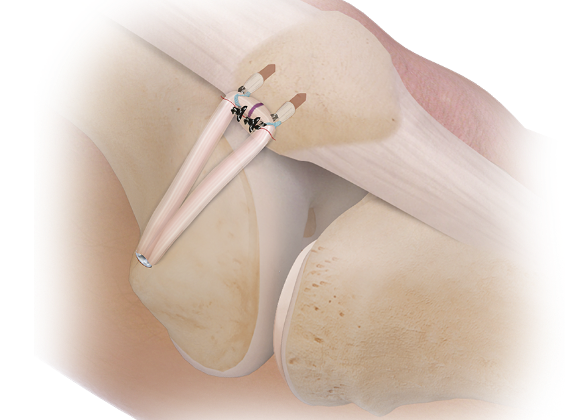
This is the foundation of surgical treatment for most recurrent instability. The torn or stretched MPFL is reconstructed using a tendon graft to recreate the main soft-tissue restraint that stops the kneecap slipping outwards. Anatomical reconstruction (placing the graft in the correct position) is more reliable than simply repairing the ligament, and MPFL reconstruction alone is often sufficient where the bony factors are mild. Dr Spelman uses either an autograft (your won tendon) or an allograft (a donor tendon) for this procedure.
Tibial tubercle osteotomy (TTO)
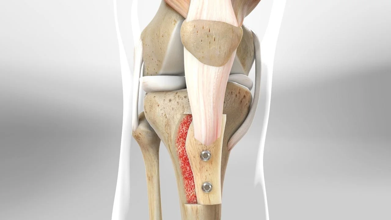
Where the bony alignment is a significant factor, the attachment of the kneecap tendon on the shin bone is repositioned. The bony attachment (tubercle) is cut and moved, then fixed with screws. It can be moved inwards (medialisation) to correct an increased TT–TG distance, downwards (distalisation) to correct a high-riding kneecap, or forwards (anteromedialisation, the Fulkerson procedure) to also offload worn cartilage on the outer/lower kneecap. A TTO is usually combined with an MPFL reconstruction.
Trochleoplasty
For high-grade trochlear dysplasia – a severely flattened or dome-shaped groove – the groove itself can be reshaped and deepened so that the kneecap sits securely. This is a more specialised procedure reserved for selected patients with severe dysplasia, and is usually combined with MPFL reconstruction.
Derotational femoral osteotomy
Where excessive inward twist of the thigh bone (increased femoral anteversion) is a major contributor, the thigh bone is cut and rotated to a more normal position, then fixed with a plate and screws. This corrects the underlying rotational cause and can also address associated valgus alignment. It is used for the subgroup of patients with significant torsional malalignment, again usually alongside an MPFL reconstruction.
Adjunctive procedures
Lateral structures that are tight can be lengthened (lateral retinacular lengthening or release), but this is used as an adjunct and is not effective as an isolated operation. Loose fragments of cartilage or bone are removed or, where suitable, fixed.
What the evidence shows
Anatomical MPFL reconstruction is reliable, with recurrent dislocation rates typically around 5% and low complication rates in most series. Combining procedures to address the bony factors improves results where those factors are significant. For severe dysplasia, trochleoplasty achieves very low redislocation rates (around 2–3% in a meta-analysis of 1,000 procedures). Derotational osteotomy improves function in patients with significant femoral torsion. The consistent theme in the evidence is that matching the operation to the underlying anatomy gives the best chance of a stable, well-functioning knee.
Complications
The risks depend on which procedure or combination is performed. Reported complications include:
General
Recurrent instability or a further dislocation despite surgery.
Stiffness (loss of movement), which may need additional physiotherapy or, occasionally, a further procedure.
Infection, blood clots (deep vein thrombosis and rarely pulmonary embolism), and persistent anterior knee pain.
MPFL reconstruction
Overall complication rates reported across series range widely (from very low up to around 30%), with recurrent instability in up to roughly 10%.
Patellar fracture is uncommon (generally under 1%) but is a recognised risk where tunnels are drilled in the kneecap; graft position that is too tight can cause pain or overload of the joint.
Tibial tubercle osteotomy
Fracture of the shin bone through the osteotomy (reported around 1–2%), delayed healing or non-union requiring re-operation, and prominent or painful screws that are commonly removed.
Trochleoplasty
Stiffness and, over the longer term, patellofemoral cartilage wear (arthritis); a proportion of patients require further surgery.
Derotational femoral osteotomy
Risks associated with cutting and healing the thigh bone – delayed union or non-union, hardware irritation, and unintended change in alignment; longer-term and return-to-sport data are more limited than for the other procedures.
Summary
Patellofemoral instability is almost always multifactorial, and lasting stability depends on identifying and addressing the specific bony, soft-tissue and rotational factors in each patient. MPFL reconstruction is the cornerstone, combined as needed with a tibial tubercle osteotomy, trochleoplasty or derotational osteotomy. Dr Spelman will assess the contributing factors in detail and tailor treatment accordingly, discussing the risks and benefits of each component with you.
MPFL reconstruction
Tibial Tubercle Osteotomy