Slipped Capital Femoral Epiphysis (SCFE)
Patient information for parents and carers — Dr Christopher Spelman, Orthopaedic Surgeon
What is SCFE
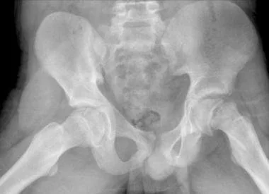
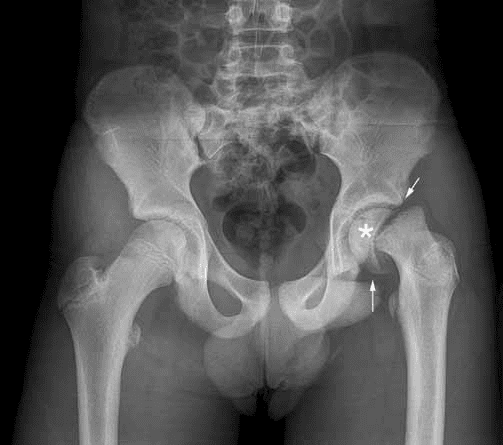
Slipped capital femoral epiphysis (SCFE) is a hip condition of older children and adolescents in which the growth plate (physis) at the top of the thigh bone weakens and the ball of the hip (the epiphysis) slips backwards and downwards relative to the neck of the femur – rather like a scoop of ice cream slipping off a cone. It most often occurs during the adolescent growth spurt, typically between about 10 and 16 years of age, and is more common in boys and in children who are above average weight. In a proportion of children an underlying hormonal (endocrine) condition contributes, and this is considered particularly in younger or atypical cases.
Symptoms
SCFE causes hip, groin, thigh or knee pain and a limp. Importantly, the pain is sometimes felt only in the knee or thigh, so the hip can be overlooked – any child or adolescent with knee or thigh pain and a limp should have their hips examined. The affected leg often turns outwards (externally rotated), may appear slightly shorter, and hip movement (particularly inward rotation) is reduced.
How SCFE is classified
Two features of the slip largely determine the treatment and the risk of complications, so they are assessed carefully:
Stability
● Stable slip – the child is able to walk, with or without crutches. These have a low risk of losing the blood supply to the ball of the hip.
● Unstable slip – the child cannot bear weight even with crutches. These behave more like an acute fracture through the growth plate and carry a much higher risk of avascular necrosis (loss of blood supply).
Severity
The slip is also graded as mild, moderate or severe according to how far the epiphysis has slipped (measured on x-ray). Greater severity increases the risk of a residual bony deformity that can cause impingement and early arthritis. The slip may also be described as acute (sudden), chronic (gradual over weeks to months) or acute-on-chronic.
Diagnosis
The diagnosis is confirmed with x-rays of both hips, including a frog-lateral view, which show the slip that may not be obvious on a standard front-on film. Because the condition can affect both hips, both are always imaged. Occasionally further imaging (MRI or CT) is used to detect a very early (pre-slip) condition or to plan complex surgery.
Why prompt treatment matters
SCFE requires urgent assessment. A child with a suspected unstable slip should not be allowed to walk on the leg and needs prompt orthopaedic review, because continued weight-bearing can worsen the slip. Treatment is almost always surgical and aims to stop any further slipping, and – depending on the severity – to correct the position of the epiphysis. The overriding goal is to protect the fragile blood supply to the ball of the hip.
Surgical treatment
The right operation depends on the stability and severity of the slip, and on surgeon and centre experience. The main options are described below. Dr Spelman will discuss which is most appropriate for your child.
Fixation in situ (pinning in situ)
This is the standard treatment for the great majority of stable, mild-to-moderate slips. Through a small incision, one screw is passed across the growth plate, under x-ray guidance, to fix the epiphysis to the neck of the femur exactly where it lies (“in situ”). It does not correct the position of the slip; instead it stabilises it to prevent any further slipping and encourages the growth plate to close. In situ fixation has a low complication rate in stable slips and preserves the blood supply well.
A single screw is used for most stable slips. A second screw adds mechanical strength (around two-thirds stiffer) and is sometimes used for unstable or severe slips, balanced against the slightly greater risk of a screw inadvertently entering the joint.
Management of the unstable slip
Unstable slips are treated urgently. They may be fixed in situ, often with gentle decompression of the joint (releasing the blood-filled swelling within the capsule) and with or without a careful, incidental repositioning of the epiphysis. The timing of surgery for unstable slips is important and somewhat controversial – some evidence suggests that operating either very early or after a delay is safer than the interval in between when a closed reduction is performed. Because the risk to the blood supply is highest in unstable slips, some centres prefer an open realignment procedure (below) that allows the blood supply to be protected directly.
Open reduction and realignment (for moderate-to-severe slips)
Where the slip is more severe and a residual deformity would be likely to cause impingement and early arthritis, the epiphysis can be repositioned to a near-anatomical position. Two main techniques are used:
● Modified Dunn procedure (subcapital realignment through a surgical hip dislocation) – the hip is safely and deliberately dislocated in a controlled way (the Ganz approach) so the surgeon can see the joint directly, shorten the femoral neck, and reposition the epiphysis while protecting the vessels that supply it. It allows the most accurate correction of the deformity. Reported avascular necrosis rates are in the order of 14% overall (around 11% in stable and 20% in unstable slips), and it is technically demanding, so it is best performed in experienced centres.
● Anterior open reduction / Fish cuneiform osteotomy (subcapital osteotomy through an anterior approach) – the epiphysis is repositioned by removing a wedge of bone from the femoral neck through the front of the hip. It also achieves realignment, with reported avascular necrosis rates of around 12–19%, and is an alternative to the modified Dunn depending on surgeon preference and experience. It has several advantages including a smaller incision and less post-operative restriction. Risk of dislocation is lower than with Modified Dunn procedure.
For a healed slip that has left a residual deformity, alternative corrective operations lower down the femur (such as a base-of-neck or intertrochanteric osteotomy) may be used, as these avoid operating directly on the fragile blood supply of the epiphysis.
The other hip (contralateral) fixation
The opposite hip develops a slip in roughly 15–35% of children, usually within the first 18 months. For this reason the other hip is monitored closely with examination and x-rays. Preventive (prophylactic) fixation of the normal-appearing other hip is considered in children at higher risk – for example, those who are younger, those with an underlying hormonal condition, or where reliable follow-up may be difficult – and this is discussed with each family.
Risks and complications
SCFE surgery is generally safe, but it carries several important risks, the most serious of which relate to the blood supply and cartilage of the hip:
● Avascular necrosis (AVN) – loss of blood supply to the ball of the hip, which can cause it to collapse and lead to early arthritis. It is the most feared complication and occurs almost exclusively with unstable slips (historically reported in around a quarter of unstable slips, and higher with some reduction techniques). It is very uncommon after in situ fixation of a stable slip.
● Chondrolysis – rapid loss of the joint cartilage causing pain and stiffness, reported in at least around 15% in some series, and sometimes associated with a screw unintentionally penetrating the joint.
● Residual deformity and femoroacetabular impingement (FAI) – a healed slip can leave a bump on the femoral neck that catches against the socket, causing pain and stiffness and sometimes requiring further surgery.
● Slip of the other hip – as above, a significant proportion of children develop a slip on the other side.
● Hardware problems – screws can be prominent, occasionally penetrate the joint, or need removal or exchange as the child grows; a further procedure is needed in a minority of patients.
● Failure to correct, or loss of position – particularly relevant to realignment procedures, and occasionally requiring revision surgery.
● General surgical risks – infection, bleeding, and the risks of anaesthesia, all of which are uncommon.
● Leg-length difference and long-term osteoarthritis – the risk of hip arthritis later in life increases with the severity of the slip (around 15–25% even for mild slips over the long term).
Post-operative protocol
The recovery plan depends on the operation performed. The following is a general guide; Dr Spelman will give your child a specific plan.
In hospital
Children usually stay in hospital for one to a few days after surgery. Pain is managed with simple medications, and a physiotherapist helps the child begin moving and using crutches before discharge.
Weight-bearing
● After in situ fixation of a stable slip, children generally use crutches with partial (protected) weight-bearing for around six weeks, progressing to full weight-bearing as comfort and healing allow.
● After an unstable slip or an open realignment procedure (modified Dunn or anterior open reduction), a longer period of protected or non-weight-bearing is usually required – often around six weeks or more – to protect the repositioned epiphysis while it heals.
Activity and return to sport
Running, jumping and sport are avoided until the growth plate has closed and healing is confirmed on x-ray – typically around three to six months, and longer after realignment surgery. Return to full activity is guided by the child’s symptoms, examination and x-rays rather than by time alone.
Follow-up
Children are reviewed regularly with x-rays to confirm that the growth plate is closing, that the screw position remains satisfactory, that avascular necrosis or chondrolysis is not developing, and to monitor the other hip. Follow-up continues until the growth plates have closed. Families are advised on the warning signs (increasing pain, stiffness or a new limp, or symptoms in the other hip) that should prompt earlier review.
Outcomes and what the evidence shows
In situ screw fixation is a reliable, safe treatment for most slips and gives excellent results for stable, mild-to-moderate slips. For severe slips, realignment procedures such as the modified Dunn can restore the anatomy and reduce impingement, with generally good hip scores, but they carry a meaningful risk of avascular necrosis, particularly in unstable slips, and are best performed in experienced centres. A systematic review comparing in situ fixation with the modified Dunn found that the Dunn achieved better correction of the bony alignment on x-ray without producing better clinical outcomes or a lower rate of avascular necrosis. For this reason, in situ fixation remains the standard for most slips, with realignment reserved for selected, more severe cases. Even with successful treatment, the long-term risk of hip arthritis increases with the severity of the original slip.
Summary
SCFE is an important adolescent hip condition that needs prompt diagnosis and, almost always, surgery. In situ screw fixation is the standard treatment and works very well for most (stable, mild-to-moderate) slips. Severe or unstable slips are more complex: they carry a higher risk of avascular necrosis and may be treated with realignment procedures such as the modified Dunn or an anterior open reduction, ideally in experienced hands. Careful post-operative rehabilitation and long-term follow-up – including of the other hip – are essential. Remember that a limping adolescent with knee or thigh pain should always have the hips examined.